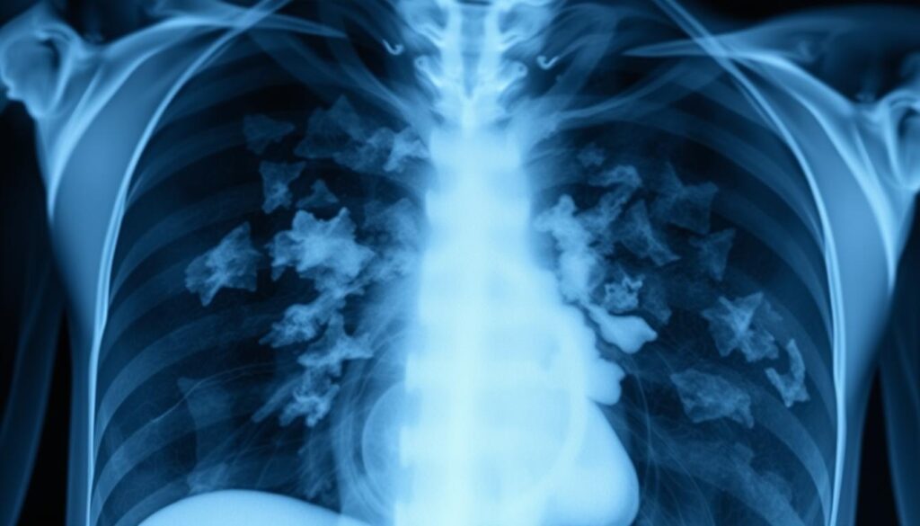
Grasping pneumonia X-ray results is vital for managing respiratory illnesses. Chest radiography helps doctors diagnose and treat lung infections effectively. Understanding these results can empower your healthcare journey1.
Chest X-rays are crucial for diagnosing pneumonia and tracking its progress. They reveal lung inflammation and infection patterns. This information guides medical professionals in creating the best treatment plan2.
Modern imaging tech has greatly improved pneumonia detection accuracy. Recent studies show precision rates of up to 98.81% in identifying lung infections2.
Key Takeaways
- Pneumonia X-rays help diagnose respiratory infections accurately
- Chest radiography reveals critical lung infection details
- Advanced imaging techniques improve diagnostic precision
- Understanding X-ray results empowers patient care
- Professional interpretation is essential for proper treatment
What is Pneumonia and Why X-Rays are Essential
Pneumonia is a serious lung disease that causes inflammation in lung tissues3. It can affect anyone, making early detection crucial. X-rays play a key role in diagnosing pneumonia and checking its severity.
Your pneumonia risk goes up if you’re in certain groups:
- Young children
- Individuals over 65
- People with existing health conditions like HIV/AIDS or lung diseases3
Spotting pneumonia signs early can speed up recovery. Common signs include:
“Early diagnosis is key to preventing serious complications” – Medical Professionals
Chest X-rays are vital for pneumonia diagnosis. They help doctors spot infections in the lungs3. These images can show white spots, pointing to infection and guiding treatment choices4.
Quick medical help can prevent dangerous outcomes like breathing problems. Your doctor might order extra tests to confirm pneumonia4. These may include blood cultures or sputum samples.
Reading Your Pneumonia X-Ray Results
X-ray results play a vital role in diagnosing and treating pneumonia. They offer detailed insights into lung health. Radiologists use these images to guide treatment plans.
Chest X-rays for pneumonia examine key aspects of lung infection. The report includes a technique section, an impression, and specific findings. These elements help doctors make informed decisions.
Normal X-Ray Appearance
A healthy chest X-ray shows clear signs of normal lung tissue4:
- Well-defined rib and diaphragm outlines
- Clear lung fields without opacity
- Symmetrical lung structures
- No visible infiltrates or consolidation
Abnormal X-Ray Findings
Pneumonia X-rays reveal specific patterns that aid diagnosis5:
| Pneumonia Type | X-Ray Characteristics |
|---|---|
| Bacterial Pneumonia | Lobar consolidation in lower lung regions |
| Viral Pneumonia | Diffuse interstitial infiltrates |
| Aspiration Pneumonia | Patchy infiltrates with consolidation |
X-ray abnormalities usually clear up after six weeks of proper antibiotic treatment5. Your doctor will use these findings to plan your care. They’ll decide if you need hospital admission or outpatient treatment4.
Quick diagnosis through X-ray interpretation can boost treatment success4. Always talk to your doctor about your X-ray results. This helps you understand the next steps in your care.
Types of Pneumonia X-Ray Patterns and Their Meaning
Chest X-rays show unique patterns for different types of pneumonia. These patterns help doctors figure out what kind of lung infection a patient has. They also show how bad the infection is.
- Bacterial Pneumonia: Characterized by lobar consolidation, which affects an entire lung lobe6
- Viral Pneumonia: Presents as diffuse interstitial infiltrates with fine, scattered linear opacities5
- Aspiration Pneumonia: Shows patchy infiltrates with potential segmental involvement7
Doctors use these patterns to identify the type of pneumonia. They also help determine how severe the infection is. Streptococcus pneumoniae causes 10-50% of community-acquired pneumonia cases worldwide7.
| Pneumonia Type | X-Ray Characteristics | Common Causes |
|---|---|---|
| Bacterial | Lobar consolidation | Streptococcus pneumoniae |
| Viral | Diffuse interstitial infiltrates | Influenza viruses |
| Aspiration | Patchy segmental infiltrates | Oral/stomach contents |
Round pneumonia often affects children. It looks like a circular spot on X-rays due to how kids’ lungs grow. As it gets worse, the spot usually becomes more solid5.
“Chest radiography is a powerful diagnostic tool that reveals the intricate details of lung infections.” – Respiratory Medicine Journal
Conclusion
Understanding your pneumonia X-ray results is vital for managing respiratory illnesses. Advanced imaging techniques have improved our ability to detect pneumonia. Chest X-ray interpretation continues to evolve with remarkable precision8.
Research shows that neural networks can now detect pneumonia with up to 94.53% accuracy9. Chest X-rays provide crucial insights into your respiratory health. Pneumonia causes over 50,000 deaths annually in the USA8.
Early detection through precise imaging can greatly improve treatment outcomes. Your doctor uses these tools to understand your pneumonia type and extent. This guides personalized treatment strategies10.
Stay proactive about your respiratory health. Regular check-ups and following medical advice are key to managing pneumonia. Medical imaging technologies continue to advance, offering sophisticated methods for monitoring respiratory illnesses9.
FAQ
What is pneumonia and how is it diagnosed?
Pneumonia is an inflammatory lung infection caused by various germs. It affects one or both lungs. Diagnosis involves physical exams, symptom evaluation, and chest X-rays.
X-rays help identify lung inflammation and specific patterns. These patterns indicate the type of pneumonia a patient might have.
What symptoms should I look for that might indicate pneumonia?
Key symptoms include persistent cough with phlegm, fever, and shortness of breath. Other signs are chills, fatigue, and chest pain.
If you experience these symptoms, consult a healthcare professional promptly. This is especially important for high-risk groups like children and older adults.
How do chest X-rays help diagnose pneumonia?
Chest X-rays confirm pneumonia by revealing lung abnormalities like infiltrates and opacities. They help differentiate between bacterial and viral pneumonia.
X-rays also assess infection severity and track treatment progress. Different pneumonia types show distinct patterns, guiding appropriate treatment.
What does a normal chest X-ray look like?
A normal chest X-ray shows clear lung fields with well-defined rib and diaphragm outlines. There are no dense patches or areas of opacity in the lung tissue.
The lungs appear uniformly dark. All anatomical structures are clearly visible and properly positioned.
What are the different types of pneumonia X-ray patterns?
Different pneumonia types present unique X-ray patterns:
– Bacterial pneumonia: Often shows lobar consolidation, affecting an entire lung lobe
– Viral pneumonia: Appears as diffuse interstitial infiltrates with fine, linear opacities
– Aspiration pneumonia: Presents patchy infiltrates with possible segmental involvement
– Round pneumonia: More common in children, appears as a round opacity
Who is at highest risk for developing pneumonia?
High-risk groups include young children, adults over 65, and people with existing health problems. Those with weakened immune systems, smokers, and individuals with prolonged hospital stays are also at risk.
People with chronic conditions like asthma, heart disease, or diabetes are more susceptible. They should take extra precautions to avoid pneumonia.
How is pneumonia typically treated?
Treatment depends on the type of pneumonia. It often includes rest, hydration, and antibiotics for bacterial pneumonia.
Viral pneumonia might require antiviral medications or supportive care. Severe cases may need hospitalization for oxygen therapy and intravenous antibiotics.
Follow-up X-rays may be used to track recovery progress. Your doctor will monitor your condition closely.
Source Links
- The diagnosis of pneumonia requires a chest radiograph (x-ray)—yes, no or sometimes? – Pneumonia – https://pneumonia.biomedcentral.com/articles/10.15172/pneu.2014.5/464
- Pneumonia detection in chest X-ray images using an ensemble of deep learning models – https://pmc.ncbi.nlm.nih.gov/articles/PMC8423280/
- Pneumonia – https://www.radiologyinfo.org/en/info/pneumonia
- Chest X-Ray For Pneumonia: Diagnosis And Interpretation – https://onestepdiagnostic.com/chest-x-ray-for-pneumonia-diagnosis/
- X-ray Atlas: Chest X-ray | GLOWM – https://www.glowm.com/atlas-page/atlasid/chestXray.html
- The Radiology Assistant : Chest X-Ray – https://radiologyassistant.nl/chest/chest-x-ray/lung-disease
- Practice Essentials, Radiography, Computed Tomography – https://emedicine.medscape.com/article/360090-overview
- A Review on Detection of Pneumonia in Chest X-ray Images Using Neural Networks – https://pmc.ncbi.nlm.nih.gov/articles/PMC9759647/
- Pneumonia detection with QCSA network on chest X-ray – Scientific Reports – https://www.nature.com/articles/s41598-023-35922-x
- Imaging of Pneumonia: An Overview – Current Radiology Reports – https://link.springer.com/article/10.1007/s40134-017-0209-9