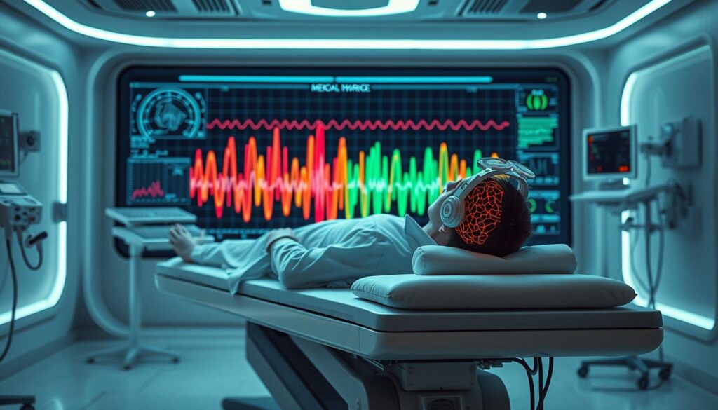
An electroencephalogram (EEG) is a cutting-edge medical test that captures your brain’s electrical activity. This non-invasive procedure helps doctors understand neural signals and diagnose neurological conditions1. Small metal discs called electrodes are placed on your scalp to measure brain activity1.
The EEG test is incredibly versatile. It helps doctors explore and diagnose various neurological disorders. These include epilepsy, seizures, sleep disorders, and brain injuries1.
An EEG usually takes about an hour to complete. However, sleep recordings might need more time1. The test is completely safe and painless1.
During the procedure, electrodes are carefully placed on your scalp. These capture the subtle electrical impulses generated by your brain cells1. No electrical current is introduced into your body during the test1.
Key Takeaways
- EEG is a non-invasive test measuring brain electrical activity
- Used to diagnose epilepsy, seizures, and sleep disorders
- Test is safe and typically takes about an hour
- Requires minimal preparation
- Helps identify various neurological conditions
Understanding EEG (Electroencephalogram) and Its Purpose
An electroencephalogram (EEG) reveals brain wave activity, offering insights into cognitive functions. This neuroimaging technique lets doctors examine your brain’s electrical patterns. EEGs provide a unique view of brain function2.
How Brain Wave Monitoring Works
EEGs measure electrical activity using electrodes on your scalp. Typically, 40 specialized electrodes capture subtle signals from your brain cells2.
Electrode caps range from 52 to 60 cm to ensure precise brain wave tracking2.
Types of Brain Wave Patterns
Your brain creates different wave patterns based on your mental state. These range from slow waves during sleep to faster ones when alert.
Neurologists use these variations to diagnose potential brain disorders3.
- Delta waves: Slowest brain waves (deep sleep)
- Theta waves: Light sleep and meditation
- Alpha waves: Relaxed wakefulness
- Beta waves: Active thinking and concentration
- Gamma waves: High-level information processing
Common Uses in Medical Diagnosis
Neurofeedback experts use EEG to diagnose and monitor various neurological conditions. This tool helps identify potential issues such as:
| Medical Condition | EEG Diagnostic Value |
|---|---|
| Epilepsy | Detecting seizure activity |
| Brain Injuries | Assessing electrical signal disruptions |
| Sleep Disorders | Analyzing brain activity during sleep stages |
| Alzheimer’s | Monitoring cognitive function changes |
EEG is a valuable diagnostic tool. However, it’s just one part of a comprehensive neurological assessment3.
“The brain is an incredible organ, and EEG helps us understand its electrical symphony.” – Neuroscience Research Team
Preparing for Your EEG Test: Essential Steps and Guidelines
Proper preparation is key for an accurate brain mapping test. Your healthcare provider will guide you through the necessary steps. Following these guidelines ensures optimal results for diagnosing neurological disorders4.
To get ready, follow these important steps:
- Wash your hair thoroughly the night before the test
- Avoid using hair conditioners or styling products
- Stop consuming caffeine 8 to 12 hours before the appointment4
- Remove hair extensions or glue-based accessories
Your medications and health are vital factors. Inform your healthcare provider about all current medications and supplements. Some drugs might affect brain wave readings4.
“Proper preparation can significantly improve the accuracy of your neurological assessment.”
On test day, wear comfy clothes and come with clean, dry hair. EEG exams can last between 45 minutes to 2 hours4.
The test is safe and painless. It’s designed to capture important brain activity information5.
These guidelines help ensure a smooth brain mapping procedure. They aid in diagnosing potential neurological disorders effectively5.
The EEG Testing Process and What to Expect
An EEG measures your brain’s electrical activity. It provides key insights into brain-computer interface interactions. This test helps doctors understand your neural signals better.
Knowing what to expect can ease your nerves. The EEG process involves specialized procedures. These procedures capture your brain’s electrical patterns accurately.
Electrode Placement and Setup
Technicians will attach 16-25 electrodes to your scalp. They use special paste or a comfortable cap. These electrodes capture neural signals from different brain areas6.
You’ll sit in a reclining chair or lie down. Staying still is important for accurate readings. The setup ensures precise data collection.
Duration and Testing Procedures
An EEG appointment typically lasts about an hour. This includes prep time and the actual recording6. Routine EEG recordings usually take 20 to 40 minutes7.
During the test, you might be asked to:
- Close your eyes and relax
- Perform deep breathing exercises
- Look at flashing lights
- Remain as still as possible
Different Types of EEG Tests
| EEG Test Type | Duration | Purpose |
|---|---|---|
| Routine EEG | 20-40 minutes | Basic brain activity assessment |
| Ambulatory EEG | 1-3 days | Continuous monitoring at home |
| Video EEG | Several days | Capture seizure activities |
Your doctor will choose the best EEG type for you. It depends on your medical needs and diagnostic requirements7. Each test serves a specific purpose.
Precision in neural signal monitoring can unlock critical insights into brain function and potential neurological conditions.
Conclusion
An EEG reveals crucial information about your brain activity. It helps doctors understand potential neurological issues. Neurologists analyze the wavy lines against established brain pattern standards8.
While not definitive for all brain disorders, EEG is a powerful diagnostic tool. It can identify conditions like brain tumors, stroke, and sleep problems9. Your doctor may suggest more tests based on initial findings8.
The brain wave monitoring technique detects subtle neural signal changes. These changes might indicate underlying health issues9. Each EEG is unique and requires expert interpretation.
EEG sensitivity varies for different neurological conditions. Some studies show detection rates of 29-55% for certain epileptic discharges8. Neurologists consider multiple factors when evaluating your results10.
An EEG is a safe, non-invasive procedure. It offers valuable insights into your neurological health. Understanding the results helps you address potential brain-related concerns effectively.
FAQ
What is an EEG test?
An EEG measures brain electrical activity using small metal electrodes on your scalp. It detects tiny charges from brain cell communication. The results appear as wavy lines on a screen or paper.
Doctors use EEGs to diagnose various neurological conditions. This non-invasive test is safe and helpful for medical professionals.
How long does an EEG test take?
A typical EEG test lasts 20 to 40 minutes. There are different types of EEG tests available. Prolonged EEGs take 1-2 hours, while ambulatory EEGs last 1-3 days at home.
Video EEGs can span several days in a hospital. The test duration depends on your specific diagnostic needs.
How should I prepare for an EEG test?
Wash your hair the night before without using conditioners or styling products. Avoid caffeine 8-12 hours before the test. Tell your doctor about all medications you’re taking.
Follow any sleep reduction instructions given. Wear comfortable clothes and arrive with clean, dry hair for the test.
Is an EEG test painful?
No, an EEG test is completely safe and painless. Electrodes are attached to your scalp using a special paste or cap. You’ll be asked to relax, close your eyes, and stay still during the procedure.
What conditions can an EEG help diagnose?
EEGs mainly diagnose epilepsy and seizure disorders. They can also identify brain tumors, damage from head injuries, and encephalopathy. EEGs help detect brain inflammation, stroke, sleep disorders, and Creutzfeldt-Jakob disease.
Additionally, they assist in diagnosing Alzheimer’s, psychoses, and narcolepsy.
What happens during an EEG test?
You’ll relax in a chair or lie on a bed with electrodes on your scalp. A technician will monitor you throughout the test. You may be asked to do tasks like deep breathing or looking at flashing lights.
The test records your brain’s electrical activity under various conditions.
Can I eat before an EEG test?
Don’t fast before the procedure, as low blood sugar might affect results. Eat a normal meal before the test. Avoid caffeine for 8-12 hours beforehand.
Follow any specific instructions from your healthcare professional about food and drink.
How are EEG results interpreted?
A neurologist analyzes the recorded brain wave patterns after the test. They compare them to normal electrical activity. The results show up as wavy lines on a chart.
A normal EEG doesn’t rule out all brain disorders. However, it provides crucial information for diagnosis and treatment planning.
Source Links
- Electroencephalogram (EEG) – https://www.healthdirect.gov.au/electroencephalogram-eeg
- Electroencephalography (EEG) and Event-Related Potentials (ERP’s) with Human Participants – https://pmc.ncbi.nlm.nih.gov/articles/PMC2909037/
- EEG: MedlinePlus Medical Encyclopedia – https://medlineplus.gov/ency/article/003931.htm
- Electroencephalogram (EEG) – https://www.hopkinsmedicine.org/health/treatment-tests-and-therapies/electroencephalogram-eeg
- EEG (electroencephalogram) – Mayo Clinic – https://www.mayoclinic.org/tests-procedures/eeg/about/pac-20393875
- Electroencephalogram (EEG) – https://www.nhs.uk/conditions/electroencephalogram/
- EEG (Electroencephalogram): Purpose, Procedure, and Risks – https://www.healthline.com/health/eeg
- Electroencephalography – https://en.wikipedia.org/wiki/Electroencephalography
- Electroencephalography During the Acute Phase Of Encephalitis: A Brief Review | Jardim Vaz de Mello – https://www.neurores.org/index.php/neurores/article/view/568/541
- Frontiers | The Effect of Electroencephalogram (EEG) Reference Choice on Information-Theoretic Measures of the Complexity and Integration of EEG Signals – https://www.frontiersin.org/journals/neuroscience/articles/10.3389/fnins.2017.00425/full