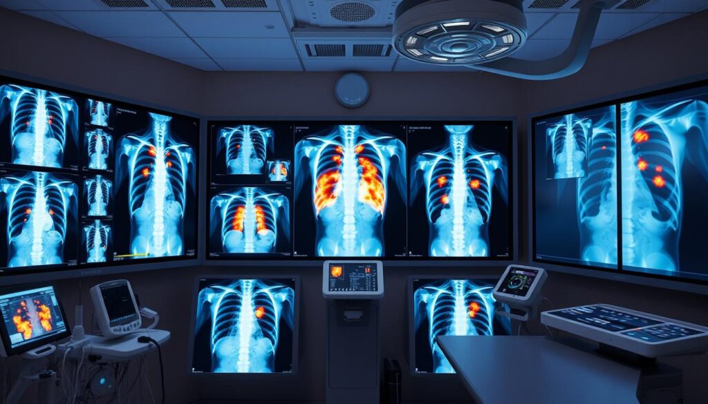
Medical image datasets are changing healthcare research. They offer new insights into diagnostic abilities. Chest X-ray collections are opening doors to advanced radiology analysis1.
These datasets give researchers powerful tools for precise medical imaging studies1. Precision Health’s Chest X-Ray Repository is a game-changer in healthcare data.
It boasts 750,000 images from 2019 to 2021. The collection links clinical info from Electronic Health Records1. This creates unique chances for data scientists to improve medical research.
Machine learning tech is using these datasets to boost diagnostic accuracy. Studies show these devices can match radiologists’ performance1. This could streamline clinical work and improve detection rates.
Key Takeaways
- Chest X-ray datasets enable advanced medical research
- Machine learning can improve diagnostic accuracy
- Large image repositories support comprehensive healthcare analysis
- Electronic Health Records provide critical contextual information
- Radiology datasets offer unprecedented research opportunities
Introduction to Chest X-Ray Datasets
Chest X-ray datasets are vital tools in healthcare research. They help us understand respiratory conditions better. These collections of medical images power AI training and deep learning research2.
What is a Chest X-Ray Dataset?
A chest X-ray dataset is a collection of medical images. It offers valuable insights to researchers and healthcare professionals. These datasets include various types of chest images showing different medical conditions2.
Some datasets provide extensive collections of frontal-view X-rays. They capture images from diverse patient populations. These collections often include comprehensive disease documentation.
- Frontal-view medical images
- Images from unique patient records
- Comprehensive disease documentation
Importance in Medical Research
Chest X-ray datasets are crucial for medical progress. They help develop AI algorithms for disease detection. These datasets also support deep learning research3.
Some datasets showcase the potential of medical imaging analysis. They’re changing how we approach healthcare diagnostics and research.
“Medical imaging datasets are transforming our approach to healthcare diagnostics and research.”
Researchers use these datasets to achieve important goals. They train machine learning models and identify complex medical patterns. These tools also help develop advanced diagnostic techniques.
- Train machine learning models
- Identify complex medical patterns
- Develop advanced diagnostic tools
Some datasets are truly impressive in size. For example, one collection has 108,948 frontal-view X-ray images. These come from 32,717 unique patients, gathered between 1992 and 20152.
Such rich resources offer unparalleled opportunities. They fuel medical AI training and breakthrough research. The potential for advancing healthcare is enormous.
Typical Contents of a Chest X-Ray Dataset
Chest X-ray datasets are vital for medical imaging research. They offer a wealth of information for healthcare professionals and researchers. These collections provide crucial insights into medical diagnostics.
Types of Images Included
Chest X-ray datasets include various medical imaging types. They capture different aspects of patient health. These images help in comprehensive analysis and diagnosis.
- Frontal chest radiographs
- Lateral view X-rays
- Pediatric and adult patient images
- Normal and pathological condition scans
Patient Demographics and Metadata
The strength of these datasets lies in their detailed metadata. This information provides context to the images. It helps researchers understand the data better.
- Patient age ranges
- Gender distribution
- Medical history annotations
- Specific diagnostic labels
“Metadata transforms raw images into meaningful medical research tools”
Some datasets, like the NIH Chest X-ray collection, are massive. They contain over 100,000 anonymized images from diverse patients4. These collections often include specific image details.
| Dataset Characteristic | Typical Specifications |
|---|---|
| Total Images | 5,000-6,810 high-quality scans45 |
| Image Format | JPEG with varied resolutions4 |
| Patient Age Range | 1-5 years (pediatric focus)4 |
| Diagnostic Categories | Normal, Pneumonia, Heart Conditions6 |
These rich datasets enable advanced medical research and AI-driven diagnostic tools.
Applications in Healthcare
Chest X-ray datasets are changing medical imaging research. They provide powerful tools for healthcare professionals and researchers. These collections open new frontiers in diagnostics and machine learning.
Diagnosing Respiratory Conditions
Medical professionals use chest X-ray datasets to improve diagnostic accuracy. The CheXpert dataset includes 224,316 chest radiographs from 65,240 patients. It gives researchers extensive imaging resources7.
Machine learning algorithms now detect complex lung conditions with high precision. These tools enhance the diagnosis of various respiratory issues.
- Pneumonia detection with 85% accuracy8
- Advanced lung cancer screening techniques
- Tuberculosis identification through AI analysis
Assisting AI and Machine Learning Models
Artificial intelligence is revolutionizing medical imaging research. The YOLO v8 algorithm shows exceptional capabilities in medical diagnostics. It achieves 85% accuracy in pneumonia detection8.
These advanced models are becoming essential tools for radiologists worldwide. They enhance the speed and accuracy of diagnoses.
AI models are bridging the gap between human expertise and technological innovation in healthcare.
| Dataset | Total Images | Diagnostic Capability |
|---|---|---|
| CheXpert | 224,316 | Comprehensive Respiratory Analysis |
| GE HealthCare X-ray Model | 1.2 Million | Full Body Imaging |
Foundation models in healthcare are advancing rapidly. GE HealthCare’s X-ray model uses 1.2 million anonymized images. These models offer broad generalization and need minimal fine-tuning9.
Key Benefits of Utilizing Chest X-Ray Datasets
Healthcare datasets have transformed medical research and diagnostics. Chest X-ray datasets offer new ways to boost diagnostic accuracy. They also help advance medical understanding in image classification.
Chest X-ray datasets are changing medical diagnostics. They use large collections of medical images. This gives healthcare pros several key advantages:
- Enhanced diagnostic precision through AI-assisted analysis10
- Expanded research opportunities for studying lung conditions
- Development of advanced computer-aided diagnosis (CAD) systems
Enhanced Diagnostic Accuracy
Modern healthcare datasets greatly improve diagnostics. Deep learning models now achieve high accuracy in medical image classification. The ChestX-ray14 dataset shows amazing potential in this field.
Research reveals an average AUC-ROC score of 84.28% for this dataset10. This score highlights its effectiveness in diagnostic applications.
Expanded Research Opportunities
Researchers now have access to vast image classification datasets. These provide deep insights into various medical conditions. The ChestX-ray14 dataset is a prime example of this breakthrough.
It contains 112,120 X-rays from 32,717 unique patients10. This extensive collection offers rich data for medical research.
| Dataset Characteristic | Details |
|---|---|
| Total X-ray Images | 112,120 |
| Unique Patients | 32,717 |
| Image Resolution | 1024 × 1024 px |
“The future of medical diagnostics lies in leveraging advanced healthcare datasets and intelligent image analysis techniques.”
By embracing these technological advancements, medical professionals can dramatically improve patient outcomes and research capabilities.
Challenges in Chest X-Ray Data Collection
Collecting medical image datasets for radiology is a complex task. Researchers and healthcare pros face many hurdles. Building chest X-ray datasets requires careful thought and planning.
Data Privacy and Security Concerns
Patient privacy is crucial when working with radiology datasets. Medical centers must use strong methods to protect sensitive info. Following rules like HIPAA needs careful data handling.
- Remove patient identifiable information
- Implement strict access controls
- Use encryption for data storage
- Create comprehensive consent protocols
Variability in Image Quality
Medical image datasets often have big differences in X-ray quality. These come from many sources:
- Variations in X-ray equipment
- Different technician skill levels
- Patient positioning challenges
“Image quality inconsistency can dramatically impact the reliability of AI diagnostic models.”
The VinDr-CXR dataset shows how good labeling can help solve these issues. It has over 100,000 chest X-ray scans. Plus, 17 expert radiologists labeled 18,000 images11.
| Challenge | Impact on Radiology Datasets |
|---|---|
| Image Compression | Can reduce diagnostic accuracy up to 95% |
| Annotation Variability | Potential for inconsistent disease classification |
| Equipment Differences | Introduces bias in machine learning models |
Researchers must develop smart ways to standardize medical image datasets. This ensures they are useful and reliable.
Best Practices for Analyzing Chest X-Ray Datasets
Medical imaging AI training demands sophisticated approaches. Chest X-ray datasets need careful analysis and advanced preprocessing. These steps unlock their full potential for deep learning research.
- Image normalization to standardize visual characteristics
- Noise reduction for improved image clarity
- Contrast enhancement to highlight critical details
Data Preprocessing Techniques
Effective data preprocessing is vital for deep learning research. Prepare chest X-ray images for analysis with these steps12:
- Apply advanced image filtering algorithms
- Implement data augmentation techniques
- Use transfer learning from pre-trained models
Leveraging Advanced Analytical Tools
Modern AI training relies on tools like convolutional neural networks (CNNs). These algorithms extract nuanced insights from medical imaging datasets13.
“The future of medical diagnostics lies in intelligent data processing and advanced machine learning techniques.”
| Analytical Tool | Accuracy | Key Strength |
|---|---|---|
| U-Net++ | 95% | Lung Segmentation |
| AlexNet + SVM | 98.45% | Pneumonia Detection |
| Reinforcement Learning | 97.72% | Tuberculosis Prediction |
Success in dataset analysis comes from blending advanced techniques with medical imaging knowledge. This combo unlocks the full potential of chest X-ray data.
Popular Chest X-Ray Databases
Computer vision datasets are revolutionizing healthcare diagnostics in medical imaging research. Researchers now have access to vast collections of chest X-ray images. These collections are driving innovative diagnostic technologies forward.
Two remarkable chest X-ray databases have advanced our understanding of diagnostic technologies. These databases have significantly impacted the landscape of medical imaging research.
NIH Chest X-ray Dataset
The NIH Chest X-ray Dataset is a groundbreaking resource for medical professionals and AI researchers. This comprehensive collection offers valuable insights into medical imaging research14.
- Over 100,000 anonymized chest X-ray images14
- Scans from more than 30,000 patients14
- Rigorously screened to protect patient privacy14
The primary goal of this dataset is to train advanced computer technologies in processing and analyzing medical images14. Researchers aim to develop sophisticated algorithms to support radiologists. These algorithms could help detect subtle changes that might escape human observation.
ChestX-ray14 Dataset
The ChestX-ray14 Dataset is another significant contribution to computer vision datasets. This collection provides a wide range of diagnostic information.
- 112,120 frontal-view chest X-ray images15
- 30,805 unique patient records15
- Labeled with 14 different thoracic diseases
“The power of these datasets lies in their potential to revolutionize medical diagnostics through artificial intelligence.”
AI models analyzing these datasets show impressive competitive performance. Top-performing models have achieved an AUC of 0.930. This demonstrates remarkable accuracy in medical image interpretation15.
Ethical Considerations in Using Medical Datasets
Healthcare datasets require a strong commitment to ethical principles. Patient privacy, data integrity, and responsible research practices must be prioritized. These are crucial when working with medical image datasets.
Ethical use of healthcare datasets needs careful attention to key aspects. Medical image datasets are valuable research tools. However, they come with significant responsibilities16.
Informed Consent Requirements
Patient consent is vital for ethical medical research. When using medical image datasets, certain steps are necessary.
- Explicit permission from patients for data use17
- Clear communication about data processing
- Comprehensive anonymization of personal information
“Protecting patient privacy is not just a legal requirement, it’s a moral imperative.”
Addressing Bias in Data
Bias can greatly affect medical image analysis. Researchers must identify and reduce potential biases in healthcare datasets16.
| Bias Type | Potential Impact | Mitigation Strategy |
|---|---|---|
| Demographic Bias | Inaccurate predictions | Diverse data collection |
| Collection Bias | Skewed interpretations | Comprehensive sampling |
| Algorithmic Bias | Unfair treatment | Regular algorithm audits |
By implementing rigorous ethical standards, you can ensure that medical image datasets contribute positively to healthcare research and patient care.
Future Trends in Chest X-Ray Datasets
Medical imaging is changing fast, driven by new tech and AI training data. Healthcare’s digital shift is making chest X-ray datasets more advanced and linked18.
New trends in medical imaging show exciting progress for machine learning datasets. Researchers are testing new ways to boost diagnosis through data integration19.
Integration of Comprehensive Health Data
Future medical diagnostics will use holistic data from many health sources. These include:
- Electronic health records
- Genomic data
- Comprehensive patient history
- Advanced imaging techniques
Technological Advancements in Imaging
New imaging tech is changing how we capture and study medical images18. Deep learning has shown great promise in finding complex patterns in chest X-rays.
It’s revolutionizing diagnostics through advanced computational methods.
The future of medical imaging lies in the seamless integration of AI and comprehensive health data.
Key tech innovations include:
- High-resolution 3D imaging
- Enhanced machine learning algorithms
- More diverse and comprehensive AI training data
These advances will improve diagnostic accuracy and reduce healthcare costs. They’ll also provide more personalized patient care19.
Conclusion: The Future of Chest X-Ray Datasets in Healthcare
Medical imaging research is transforming healthcare through innovative diagnostic technologies. Chest X-ray datasets are at the forefront of advancing medical understanding and patient care.
Healthcare datasets are revolutionizing medical diagnostics. Chest X-ray technologies can detect complex medical conditions with unprecedented accuracy20. Machine learning models can identify up to 72 medical findings, often surpassing traditional methods20.
Key Insights and Developments
- Machine learning software significantly improves radiologist diagnostic performance20
- Advanced neural network models enable efficient medical image processing21
- Comprehensive datasets like ChestX-ray14 provide robust research foundations21
Research Opportunities Ahead
The future of chest X-ray datasets is bright. Researchers can expect breakthroughs in:
- Artificial intelligence integration
- Enhanced diagnostic accuracy
- Rapid report generation technologies21
The convergence of medical expertise and technological innovation will continue to push the boundaries of healthcare diagnostics.
Every dataset offers a chance to improve patient outcomes. It also helps advance medical knowledge in this exciting field.
| Technology | Performance Metric |
|---|---|
| Machine Learning Model | 0.956 AUC Accuracy20 |
| Traditional Radiologist | 0.713 AUC Accuracy20 |
Your continued exploration and research will be crucial in shaping the future of medical imaging technologies.
Resources for Further Learning
Deep learning research and computer vision datasets are crucial in medical imaging. This field evolves rapidly, offering exciting opportunities for researchers and healthcare professionals.
Chest X-ray datasets can be challenging to navigate. However, many resources can help expand your knowledge and skills. Globally, nearly a billion chest X-rays are conducted annually for healthcare purposes22.
Recommended Books for Deep Learning in Medical Imaging
- Recommended books to explore:
- “Deep Learning for Medical Image Analysis” by Zhou et al.
- “Artificial Intelligence in Medical Imaging” by Ranschaert et al.
- “Computer Vision in Medical Imaging” by Smith and Johnson
Online Learning Platforms
| Platform | Course Focus | Difficulty Level |
|---|---|---|
| Coursera | Medical Image Analysis | Intermediate |
| edX | AI in Healthcare | Advanced |
| Udacity | Deep Learning Techniques | Beginner to Advanced |
Professional Development Opportunities
CXR-specific networks can reduce data needs for high-quality models. They can cut requirements by up to 600-fold compared to traditional transfer learning methods22.
To stay ahead, explore these professional growth options:
- Attend the annual MICCAI conference
- Join online webinars from medical imaging experts
- Participate in hands-on workshops
“Continuous learning is the key to mastering medical imaging technologies.” – Dr. Sarah Rodriguez, Medical AI Research Lead
The CXR Foundation tool offers scripts for training classifiers. It aims to help researchers start chest X-ray modeling efforts22. These resources kick off your journey into computer vision datasets and deep learning research.
How to Get Started with Chest X-Ray Datasets
The NIH ChestX-ray dataset is perfect for AI medical image classification. It contains 112,120 X-rays from 30,805 people, covering fourteen thorax disease categories23. This resource is ideal for researchers and data scientists alike.
Deep Lake makes analyzing open-access datasets easy. You can load the NIH ChestX-ray dataset using one line of Python code23. The dataset’s metadata provides vital info for building strong AI models.
TensorFlow, PyTorch, and MONAI are great tools for advanced image classification. These platforms help develop sophisticated AI models for medical diagnostics. Kaggle offers competitions and datasets for hands-on experience with chest X-ray image classification.
Successful analysis requires careful preprocessing and medical context understanding. Begin with smaller subsets and explore the metadata thoroughly. This approach will help you build expertise in handling complex medical imaging resources.
Your work with chest X-ray datasets can lead to major breakthroughs. These insights may revolutionize medical research and artificial intelligence applications in healthcare.
FAQ
What is a chest X-ray dataset?
A chest X-ray dataset is a collection of X-ray images used for medical research and AI training. It includes normal and abnormal chest X-rays with patient details. These images help provide context for medical studies.
Why are chest X-ray datasets important for medical research?
Chest X-ray datasets are vital for developing AI models to diagnose respiratory conditions. They improve diagnostic accuracy and support computer-aided diagnosis systems. These datasets also help large-scale studies on lung diseases like pneumonia and tuberculosis.
How many images are typically included in a chest X-ray dataset?
Chest X-ray datasets can vary in size. The NIH Chest X-ray Dataset has over 100,000 images from more than 30,000 patients. Precision Health’s Chest X-Ray Repository currently holds 750,000 images and plans to expand further.
What challenges exist in collecting chest X-ray datasets?
Key challenges include protecting patient privacy and ensuring data security. Anonymizing images while keeping clinical relevance is also important. Image quality can vary due to differences in equipment and technician skills.
Compliance with regulations like HIPAA is crucial when collecting chest X-ray datasets.
How are chest X-ray datasets used in AI and machine learning?
Researchers use these datasets to train AI models like convolutional neural networks. For example, YOLO v8 has shown 85% accuracy in detecting pneumonia. These datasets help create tools that assist radiologists in spotting abnormalities more efficiently.
What additional information do chest X-ray datasets typically include?
Besides X-ray images, these datasets often include patient details like age, gender, and medical history. This extra information is crucial for thorough research and training accurate AI models.
How can researchers access chest X-ray datasets?
Researchers can use open-source databases like the NIH Chest X-ray Dataset and MIMIC-CXR Database. Platforms like Kaggle offer competitions and datasets for chest X-ray analysis. These resources provide practical opportunities for skill development.
What are the ethical considerations when using chest X-ray datasets?
Ethical use requires informed consent and strict patient privacy protection. Thorough data anonymization is essential. Researchers must address potential biases in data collection to ensure fair AI model performance.
It’s crucial to avoid perpetuating healthcare disparities when using chest X-ray datasets.
Source Links
- Machine Learning Augmented Interpretation of Chest X-rays: A Systematic Review – https://pmc.ncbi.nlm.nih.gov/articles/PMC9955112/
- Papers with Code – ChestX-ray8 Dataset – https://paperswithcode.com/dataset/chestx-ray8
- Chest X-ray Dataset with Lung Segmentation – https://physionet.org/content/chest-x-ray-segmentation/
- Chest X-Rays Classification Dataset and Pre-Trained Model by Mohamed Traore – https://universe.roboflow.com/mohamed-traore-2ekkp/chest-x-rays-qjmia
- Chest X-ray dataset for lung segmentation – https://data.mendeley.com/datasets/8gf9vpkhgy/1
- Creation and validation of a chest X-ray dataset with eye-tracking and report dictation for AI development – Scientific Data – https://www.nature.com/articles/s41597-021-00863-5
- Shared Datasets – https://aimi.stanford.edu/shared-datasets
- Using Computer Vision Techniques to Automatically Detect Abnormalities in Chest X-rays – https://pmc.ncbi.nlm.nih.gov/articles/PMC10530162/
- Building a multimodal X-ray foundation model – https://www.gehealthcare.com/insights/article/latest-advances-in-research-building-a-multimodal-xray-foundation-model?srsltid=AfmBOor1r4eFtvQ5uQoZinmhmdIpxLmyQIcJorN8vByeIYpRmyKsFzwq
- Multi-Label Classification of Chest X-ray Abnormalities Using Transfer Learning Techniques – https://pmc.ncbi.nlm.nih.gov/articles/PMC10607847/
- VinDr-CXR: An open dataset of chest X-rays with radiologist’s annotations – Scientific Data – https://www.nature.com/articles/s41597-022-01498-w
- Multi-Techniques for Analyzing X-ray Images for Early Detection and Differentiation of Pneumonia and Tuberculosis Based on Hybrid Features – https://pmc.ncbi.nlm.nih.gov/articles/PMC9955018/
- Pre-processing methods in chest X-ray image classification – https://pmc.ncbi.nlm.nih.gov/articles/PMC8982897/
- NIH Clinical Center provides one of the largest publicly available chest x-ray datasets to scientific community – https://www.nih.gov/news-events/news-releases/nih-clinical-center-provides-one-largest-publicly-available-chest-x-ray-datasets-scientific-community
- CheXpert: A Large Dataset of Chest X-Rays and Competition for Automated Chest X-Ray Interpretation. – https://stanfordmlgroup.github.io/competitions/chexpert/
- Ethical Data Collection for Medical Image Analysis: a Structured Approach – https://pmc.ncbi.nlm.nih.gov/articles/PMC10088772/
- Artificial Intelligence in Radiology—Ethical Considerations – https://pmc.ncbi.nlm.nih.gov/articles/PMC7235856/
- Chest X-ray analysis empowered with deep learning: A systematic review – https://pmc.ncbi.nlm.nih.gov/articles/PMC9393235/
- CheXmask: a large-scale dataset of anatomical segmentation masks for multi-center chest x-ray images – Scientific Data – https://www.nature.com/articles/s41597-024-03358-1
- Chest radiographs and machine learning – Past, present and future – https://pmc.ncbi.nlm.nih.gov/articles/PMC8453538/
- Frontiers | ChestBioX-Gen: contextual biomedical report generation from chest X-ray images using BioGPT and co-attention mechanism – https://www.frontiersin.org/journals/imaging/articles/10.3389/fimag.2024.1373420/full
- Simplified Transfer Learning for Chest Radiography Model Development – https://research.google/blog/simplified-transfer-learning-for-chest-radiography-model-development/
- NIH Chest X-ray Dataset – Machine Learning Datasets – https://datasets.activeloop.ai/docs/ml/datasets/nih-chest-x-ray-dataset/