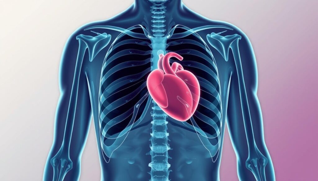
Chest X-rays are vital for understanding your chest cavity’s health. This imaging method uses low-dose radiation to create detailed pictures of your internal structures1. Your doctor might suggest a chest X-ray to check for heart, lung, or tissue issues1.
Radiographers take images of key areas during pulmonary diagnosis. These include your heart, lungs, bronchi, and chest bones1. The process is quick and easy, with little prep needed2.
Chest X-rays are done in various medical settings. They’re common in emergency rooms and regular doctor’s offices2. While there’s some radiation exposure, doctors believe the benefits outweigh potential risks2.
Key Takeaways
- Chest X-rays provide detailed internal imaging of thoracic structures
- The procedure is quick, non-invasive, and widely available
- Radiation exposure is minimal and carefully monitored
- Digital technology allows rapid result interpretation
- Used for diagnosing multiple health conditions
Understanding Chest X-Ray Basics and Benefits
Chest X-rays are vital for modern medical diagnostics. They offer a view into your body’s internal structures. These tools help doctors detect and diagnose various conditions quickly.
Chest X-rays are the most common diagnostic imaging exam. They provide key insights into lung and heart health3. Doctors use them in Lung Disease Detection and Cardiothoracic Radiology to spot health issues4.
What is a Chest X-Ray?
A chest X-ray is a safe medical test. It captures black-and-white images of your chest’s internal structures. Low-radiation X-ray beams pass through your body during the procedure.
This creates detailed images of your chest. Different tissues absorb radiation differently, making the images clear4.
Key Benefits and Applications
- Rapid Pneumonia Screening
- Tuberculosis Screening
- Evaluation of heart and lung conditions
- Detection of fractures and medical device placement4
Common Conditions Detected
Chest X-rays can reveal many medical conditions, including:
- Pneumonia
- Heart failure
- Lung cancer
- Emphysema
- Calcium deposits in blood vessels3
The procedure typically takes about 15 minutes and provides invaluable diagnostic information3.
Safety note: Chest X-rays use very low radiation. It’s often lower than natural radiation sources4. Tell your technician if you’re pregnant for proper precautions.
Preparing for Your Chest X-Ray Procedure
Chest X-rays are vital for assessing respiratory health. A few simple steps can ensure accurate results. Your cooperation will make the process smooth and efficient5.
- Remove clothing from the waist up
- Change into a hospital gown
- Take off jewelry or metal objects from the chest area
- Inform your healthcare provider about potential pregnancy
The chest X-ray procedure involves several steps:
- Stand against the imaging plate
- Position your arms up or to the sides
- Roll your shoulders forward
- Take a deep breath and hold it for a few seconds
“A well-prepared patient ensures the most accurate diagnostic imaging.” – Medical Imaging Professionals
X-rays are quick and painless. They can spot many health issues, like bone fractures and lung problems. The radiation exposure is low and safe for diagnostic use65.
| Preparation Step | Purpose |
|---|---|
| Remove metal objects | Prevent image interference |
| Wear hospital gown | Ensure clear chest imaging |
| Follow positioning instructions | Obtain comprehensive view |
Pro tip: You can usually resume normal activities right after the X-ray5.
Conclusion
Chest X-ray radiography provides vital insights into your pulmonary health. A chest X-ray can reveal hidden health conditions. It helps doctors identify heart valve issues and lung problems7.
Your safety is a top priority during the procedure. Chest X-rays use minimal radiation, making them a safe diagnostic tool. Radiologists carefully examine each image for accurate interpretation7.
The process is quick and simple. It usually involves taking images from two angles. This gives a complete view of your chest structures.
Chest X-rays are powerful but just one part of your health journey. Talk openly with your doctor about any concerns. If more tests are needed, see them as steps to better health.
Your commitment to thorough diagnosis can make a big difference. It can lead to early detection and better treatment. Stay proactive in understanding and maintaining your health.
FAQ
What exactly is a chest X-ray?
A chest X-ray is a medical imaging test. It uses low-dose radiation to create detailed pictures of your chest’s internal structures. These images help healthcare providers diagnose and monitor various medical conditions.
The X-ray shows different tissue densities in black, white, and gray. It reveals your heart, lungs, blood vessels, airways, and chest bones.
How long does a chest X-ray procedure take?
The chest X-ray procedure is typically very quick. It usually lasts only about 10-15 minutes. The actual imaging takes just a few seconds.
Most of the time is spent positioning you correctly. This ensures clear images are captured.
Is a chest X-ray safe?
Chest X-rays are generally considered very safe. They involve minimal radiation exposure. The amount is comparable to a few days of natural background radiation.
However, pregnant women should inform their healthcare provider before the procedure. This precaution ensures the safety of both mother and baby.
What should I wear for a chest X-ray?
You’ll be asked to remove clothing from the waist up. A hospital gown will be provided for you to wear. Remove any jewelry or metal objects from your chest area.
These items can interfere with the X-ray images. Wear comfortable, loose-fitting clothing that’s easy to remove.
What conditions can a chest X-ray detect?
Chest X-rays can detect various conditions. These include pneumonia, lung infections, and congestive heart failure. They can also spot lung cancer and broken ribs or chest bones.
X-rays reveal an enlarged heart or fluid around the lungs. They show the placement of medical devices like pacemakers or catheters.
Do I need to prepare special for a chest X-ray?
Generally, no special preparation is required. You don’t need to fast or change your diet or medication. Just inform your healthcare provider if you’re pregnant or might be.
Be prepared to remove upper body clothing and jewelry. That’s all the preparation you need.
Will the chest X-ray procedure hurt?
The chest X-ray is completely painless. You’ll be asked to stand still and hold certain positions. You might need to hold your breath briefly during imaging.
The entire process is quick. It involves no invasive procedures, ensuring your comfort throughout.
How soon will I get my chest X-ray results?
Result times can vary depending on your healthcare facility. Some providers can review images immediately. Others might take a few days.
A radiologist will analyze the images. They’ll send a detailed report to your healthcare provider. Your provider will then discuss the findings with you.
Source Links
- Chest X-Ray – https://www.hopkinsmedicine.org/health/treatment-tests-and-therapies/chest-xray
- X-ray: Imaging test quickly helps find diagnosis – https://www.mayoclinic.org/tests-procedures/x-ray/about/pac-20395303
- chestrad – https://www.radiologyinfo.org/en/info/chestrad
- Chest X-rays – Mayo Clinic – https://www.mayoclinic.org/tests-procedures/chest-x-rays/about/pac-20393494
- X-ray – https://www.nhs.uk/conditions/x-ray/
- Chest X-Ray: Purpose, Procedure, and Risks – https://www.healthline.com/health/chest-x-ray
- What Do My Chest X-Ray Results Show? – Baptist Health – https://www.baptisthealth.com/blog/heart-care/what-do-my-chest-x-ray-results-show